Ультразвуковое исследование первых плюснефаланговых суставов на тофусы и голеностопных суставов на признак двойного может быть рекомендовано в качестве скринингового для раннего выявления подагры по УЗИ в повседневной практике.

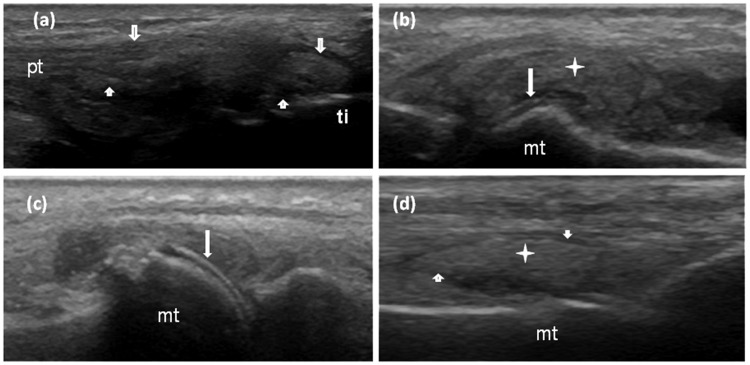
Цель. Определить оптимальные места для выявления подагры на ранней стадии методом ультразвукового исследования. Методы. Была проведена проспективная оценка шестидесяти пациентов с диагнозом подагры, поставленным по наличию кристаллов моноурата натрия (25 в раннем периоде [симптомы наблюдаются ≤ 2 лет], 35 в позднем периоде [симптомы наблюдаются> 2 лет]) и 36 здоровых добровольцев с нормоурикемией для контроля. Для выявления тофусов и феномена двойного контура проводили стандартизованное маскированное ультразвуковое исследование (УЗИ) 36 суставов, трицепсов и коленных сухожилий. Результаты. В раннем периоде чувствительность УЗИ была ниже, чем в позднем. Бинарный логистический регрессионный анализ показал, что два признака на УЗИ (тофус в первом плюснефаланговом суставе стопы [отношение шансов = 16,46] и феномен ДК в голеностопном суставе [отношение шансов = 25,18]) вносили значимый вклад в конечную модель ранней диагностики подагры (чувствительность и специфичность составили 84% и 81%, соответственно). Значение каппа надежности оценок между экспертами для феномена ДК и тофуса составило 0,712. Выводы. Обследование четырех суставов (обоих первых плюснефаланговых суставов на тофусы и обоих голеностопных суставов на признак ДК) легко осуществимо и надежно. Такое обследование можно использовать в качестве скринингового для раннего выявления подагры по УЗИ в повседневной практике.
Norkuviene E, Petraitis M, Apanaviciene I, Virviciute D, Baranauskaite A.
Journal of International Medical Research. 2017 Jan 1:300060517706800.
PMID: 28617199
PMCID: PMC5625526
DOI: 10.1177/0300060517706800
Спонсор выпуска новостей
An optimal ultrasonographic diagnostic test for early gout: A prospective controlled study.
Abstract
Objective To identify the optimal sites for classification of early gout by ultrasonography. Methods Sixty patients with monosodium urate crystal-proven gout (25 with early gout [≤2-year symptom duration], 35 with late gout [>2-year symptom duration], and 36 normouricemic healthy controls) from one centre were prospectively evaluated. Standardized blinded ultrasound examination of 36 joints and the triceps and patellar tendons was performed to identify tophi and the double contour (DC) sign. Results Ultrasonographic sensitivity was lower in early than late gout. Binary logistic regression analysis showed that two ultrasonographic signs (tophi in the first metatarsophalangeal joint [odds ratio, 16.46] and the DC sign in the ankle [odds ratio, 25.18]) significantly contributed to the final model for early gout diagnosis (sensitivity and specificity of 84% and 81%, respectively). The inter-reader reliability kappa value for the DC sign and tophi was 0.712. Conclusions Four-joint investigation (both first metatarsophalangeal joints for tophi and both ankles for the DC sign) is feasible and reliable and could be proposed as a screening test for early ultrasonographic gout classification in daily practice.
KEYWORDS:
Gout; diagnostic test; double contour sign; tophus; ultrasound


